Problem
Bone tissue continuously remodels throughout life, adapting to factors such as age, hormonal status, and mechanical loading. These same factors also shape how bone heals after injury or implantation of biomaterials. Despite their importance, patient-specific characteristics such as sex and age are rarely correlated directly to histological images, leaving potential biological differences underexplored.
This project aims to reveal how bone microarchitecture varies across individuals by using artificial intelligence. Specifically, we will train AI models to predict patient sex and age directly from whole-slide images (WSIs) of human bone. Beyond the predictive task itself, the focus is on understanding which microscopic features drive these predictions - offering a new window into the biological signals encoded in digital pathology data.
Approach
You will develop a deep learning pipeline that links whole-slide histology with patient metadata using multiple instance learning (MIL). The project has two main components:
- Model development: Train and validate a MIL model to predict slide-level sex and age from WSIs using only slide-level labels.
- Interpretability and quantification: Derive attention heatmaps to identify which tissue regions guide model predictions, and link these regions to quantitative histological features (e.g., bone density).
Through these steps, you will advance a framework that combines explainable AI with computational histomorphometry, helping uncover how age- and sex-related features manifest in bone tissue.
Data
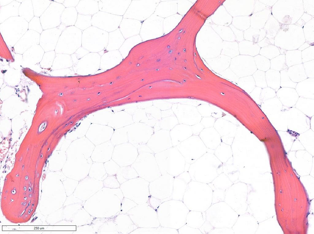
The dataset consists of more than 300 anonymized human bone whole-slide images, each paired with metadata including patient sex and age. Slides are H&E-stained and digitized at high resolution. Tissue regions are curated to include both cortical and trabecular bone compartments, enabling robust model training and interpretability analysis.
Requirements
- Master’s student in Artificial Intelligence, Computer Science, Biomedical Engineering, or a related field.
- Strong programming skills in Python, with experience in PyTorch or another deep learning framework.
- Affinity with medical image analysis, explainable AI, or multiple instance learning.
- Interest in applying AI to real-world biomedical problems; no wet-lab work required.
Information
- Project duration: 6–9 months
- Location: Radboud University Medical Center, Nijmegen, The Netherlands
- You will join the Computational Pathology Group, a leading research team for deep learning in digital pathology. The project is supervised by Gerry Koons, Marie Skłodowska-Curie Postdoctoral Fellow, and will provide access to a large GPU cluster, high-resolution pathology data, and interdisciplinary mentorship bridging AI, pathology, and regenerative medicine.
- For more information, please contact Gerry Koons.