Musculoskeletal image analysis

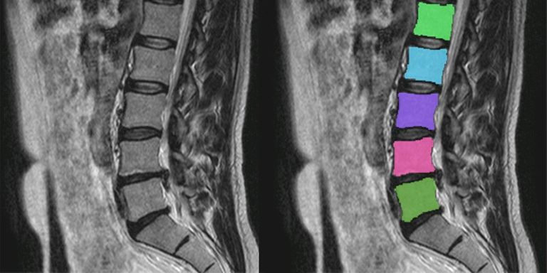
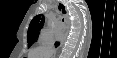
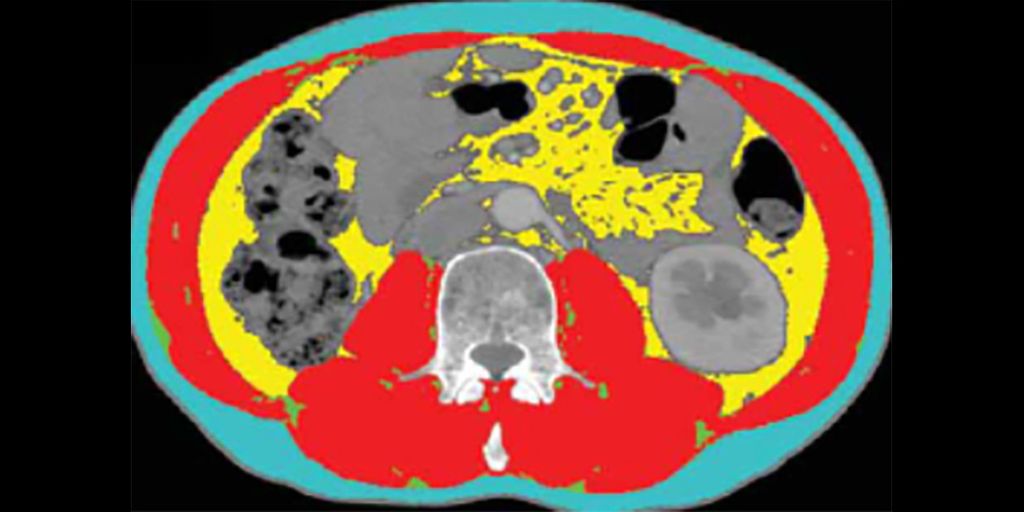
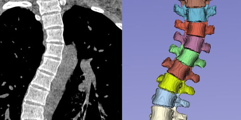
Musculoskeletal diseases are responsible for more years lived with disability than any other kind of chronic condition. We develop image analysis algorithms that assist radiologists and orthopedic surgeons in recognizing musculoskeletal conditions early and in making well-informed treatment decisions based on quantitative imaging data. This research line was led by Nikolas Lessmann.

Funding

EFRO Oost-Nederland

Projects

MRI-based decision support tool for patients with chronic lower back pain

We develop AI-based MRI analysis software for treatment decision support in patients with chronic degenerative low back pain.

Read more →
Detecting and characterizing vertebral fractures in CT scans

Developing image analysis algorithms that automatically detect osteoporotic vertebral fractures.

Read more →
Body composition assessment in 3D CT and MR images

Automatic quantification of muscle and fat tissue in 3D CT and MR images

Read more →
Scoliosis simulation for improving a segmentation and labelling algorithm

Modeling of deformities in adolescent idiopathic scoliosis to improve segmentation

Read more →

Algorithms

Several algorithms that this group has developed can be tried on the grand-challenge.org platform:

Vertebra segmentation and labeling

Segmentation and labeling of the vertebrae in CT scans with arbitrary field of view.

Vertebral Fracture Assessment

A neural network that assesses vertebral fractures according to the Genant classification

Vertebral Abnormality Scoring

Score from 0 to 100 that expresses how abnormal the shape of a vertebra is

Rib segmentation

Segments and labels the ribs in CT images

Femur segmentation in CT

Segments the left and right femur in CT images

Hip segmentation in CT

Segments the left and right hip bones in CT images

Tibia segmentation in CT

Segments the left and right tibia in CT images

Wrist segmentation

Segments the carpal bones and radius and ulna in (dynamic) CT scans of the wrist.

Body composition

Locates L3 in a CT scan and measures the area of subcutaneous, visceral, and intermuscular adipose tissue and smooth muscle. The average Hounsfield Units of each area are also computed.

People

Nikolas Lessmann

Nikolas Lessmann

Assistant Professor

Matthieu Rutten

Matthieu Rutten

Associate Professor, Radiologist

Lena Philipp

Lena Philipp

PhD Candidate

Lars Leijten

Lars Leijten

PhD Candidate