Clinical Problem
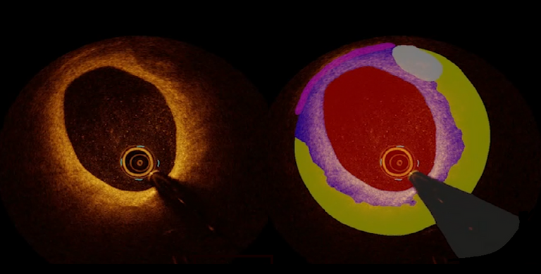
Intracoronary optical coherence tomography (OCT) is a powerful imaging technique that allows for high-resolution visualization of coronary artery structures. Intracoronary OCT is increasingly used during cardiac interventions, improving the results of cardiac procedures for patients with myocardial infarction. However, the interpretation of OCT images is challenging due to the high resolution nature of OCT-data, as well as the complexity and variability of coronary plaques and other vascular structures.
To enable automated and standardized plaque characterization, the CARA lab (https://www.cara-ai-lab.nl) is developing deep learning-based models for accurate segmentation of these structures. The current model, OCT-AID, is designed to segment important vessel wall structures and various plaque types, but there is room for improvement to enhance its diagnostic and prognostic capabilities.
The goal of this project is to improve the existing algorithm package, by expanding the current segmentation model and include additional structures, not only focusing on plaque types but also on inflammatory features such as macrophages and cholesterol crystals. Other aims are testing state-of-the-art AI architectures to improve model accuracy and predictions speed. By doing so, we aim to increase the diagnostic and prognostic value of OCT images, facilitating better clinical decision-making. Additionally, we aim to validate our models on external datasets, ensuring that the model is more generalizable and ready for real-world clinical use.
Objectives / Aims / Goals
Primary Objective:
To enhance the current multiclass segmentation model by integrating new classes of structures, and testing and validating its performance for increased diagnostic and prognostic accuracy.
Further goals (depending on project scope, student interests, literature findings, and initial conclusions):
- Develop an improved model for segmentation of specific structures
- Develop datasets for internal and external evaluation of the models
- (Re)train models on diverse and/or more challenging datasets
- Investigate clinical usability of the model at the cardiac catheterization lab
- Conduct subgroup analyses to assess model performance across different patient populations
- Ensure all model outputs can be integrated into our AI pipelines
Requirements
- Master students with a major in computer science, artificial intelligence, mathematics, biomedical engineering, technical medicine, physics, or a related area in the final stage of master's studies
- Experience with programming in Python
- Experience in deep learning, medical imaging, and/or medical image analysis
Practical Information
- Project Duration: Variable (6 months for MSc thesis)
- Location: CARA Lab, Department of Cardiology, Radboud University Medical Center, Nijmegen
- For more information, please contact Jos Thannhauser, Ruben van der Waerden or Thijs Luttikholt.
Suggested Reading
- Volleberg R, Waerden van der R, et al., "Comprehensive full-vessel segmentation and volumetric plaque quantification for intracoronary optical coherence tomography using deep learning", Mar 2025. doi: [https://doi.org/10.1093/ehjdh/ztaf021]