Background
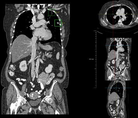
Patients with cancer typically undergo many imaging exams, mostly with computed tomography (CT). First, CT imaging is used for diagnosis and staging. During and after treatment, CT imaging is used to monitor the progression of the disease. Radiologists need to carefully go over these CT scans, and compare them with the prior CT scans of the same patient. In this project we focus on thorax-abdomen CT scans, as these are the most often acquired and these scans can contain up to 1000 slices and it can take up to an hour to carefully inspect and report them.
The Diagnostic Image Analysis Group has started a large project to develop AI tools to help speed up and improve the reporting of CT thorax-abdomen scans of oncology patients. Two PhD students have recently started. One student focuses on registration of a CT scan with the prior scans of the same patient, producing subtraction images, and detect clinically relevant changes from these series of exams. The other student focuses on the detection and quantification of bone metastases. In this third project, we specifically want to focus on the detection and quantification of abnormal lymph nodes and other lesions in the abdomen.
This project is funded by Eurostars and is a collaboration with Thirona and Merantix. You will be supervised by Bram van Ginneken.
Requirements
You should be a creative and enthusiastic researcher with an MSc degree in Computer Science, Physics, Engineering or Biomedical Sciences or similar, with a clear interest to develop image analysis algorithms and an affinity with medical topics. Experience with deep learning, machine learning, and image analysis is a plus. Good communication skills and expertise in software development, preferably in Python, are essential.
Terms of employment
You will be appointed for four years as a PhD student with the standard salary and secondary conditions for PhD students in the Netherlands. The research should result in a PhD thesis.
Organization
You will work in the Diagnostic Image Analysis Group (DIAG) DIAG is part of the Departments of Imaging, Pathology, Ophthalmology, and Radiation Oncology of Radboud University Medical Center. We develop computer algorithms to interpret and process medical images. The group currently consists of around 70 researchers. Radboud University Medical Center and Radboud University are located in Nijmegen, the oldest Dutch city with a rich history and one of the liveliest city centers in the Netherlands. Radboud University has over 17,000 students. Radboud UMC is a leading academic center for medical science, education and health care with over 8,500 staff and 3,000 students.
Information
For more information please contact Bram van Ginneken by e-mail.
Application
You can send applications as a single pdf file to bram.vanginneken@radboudumc.nl. In this pdf file the following should be included: CV, list of followed courses and grades, letter of motivation, and preferably a reprint of your Master thesis or any publications in English you have written. This application will remain open until the position has been filled. Applications are processed immediately upon receipt.
