This is an AI for Health MSc project. Students are elgible to receive a monthly reimbursement of €500,- for a period of six months. For more information please read the requirements.
Clinical problem
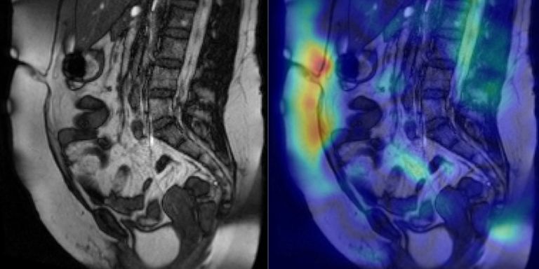
This project aims to improve outcomes in the 30,000 patients per year in the Netherlands that develop chronic pain after abdominal surgery. This pain is often caused by adhesions, tough bands of tissue that form between structures and organs in the abdomen. Medical consumption in these patients is high, 70% is under treatment of a gastroenterologist and one in three is permanently taking pain suppressing medication. Adhesions can be detected both invasively and non-invasively. Invasive diagnosis (i.e. surgery) is controversial, because it can cause the formation of new adhesions. Non-invasively, adhesions are detected with either ultrasound or cine MRI. Ultrasound works well, but can only detect adhesions attached to the front abdominal wall. Cine MRI, on the other hand, can detect adhesions in the entire abdomen. This type of imaging is used in the Radboudumc to make treatment decisions for patients with chronic abdominal pain. It helps doctors decide whether a patient should be treated conservatively with medication, or more progressively with surgery. Cine MRI is not widely used in other hospitals yet, because radiological reading is time-consuming and expertise-dependent.
Automatic detection algorithms for adhesion detection can reduce the learning curve for new readers and reduce variability among radiologists. By helping radiologists interpret the images, the algorithm may reduce the number of false positives and negatives. False positives can result in unnecessary surgery, whereas false negatives can block surgery for patients who may actually be helped by it.
The gif below gives an impression of the type of data the student will be working with.
Solution
We want to develop an automated system for adhesion detection, using deep learning segmentation and classification. We take an existing, semi-automated method as a starting point. This existing method need a segmentation map of the abdominal cavity, to perform a masked image registration. This allows quantification of the amount and direction of movement of the abdomen and its contents. The local difference in movement between the abdominal wall and its contents, called visceral slide, is indicative for adhesions. Little or no visceral slide indicates that an adhesion is present. This method can be fully automated using deep learning-based segmentation of the abdominal cavity and deep learning-based classification or detection on the registration results.
If the automation of this existing method is finished quickly, the student is encouraged to develop other (deep learning) approaches. Adhesion detection is new and unexplored territory in AI research and leaves a lot of room for creative and challenging approaches!
Data
The student will have immediate access to 60 cine MRI studies with binary labels (adhesions or no adhesions) extracted from radiology reports and abdominal cavity segmentations. In 10 of these studies, adhesions are annotated with bounding boxes. Soon, this database will be extended to more than 300 cine MRI studies with labels from another hospital.
Approach
Students will be supervised by a DIAG research member whose research is dedicated to adhesion detection with machine learning techniques. Depending on the project progression, they will be guided to set up an automatic CNN-based system for detecting adhesions to the abdominal wall, in the lesser pelvis and between bowel loops. The final deliverable will be a publicly available algorithm on the platform Grand Challenge. Students get access to the high-performance deep learning cluster of DIAG, named SOL, for running machine learning experiments.
Requirements
- Student with a major in data science, computer science, or artificial intelligence in the final stage of master level studies
- Interest in (medical) image analysis and machine learning
- Affinity with programming in Python and with deep learning packages (e.g. PyTorch) is required
Information
- Project duration: 6 months
- Location: Radboud University Medical Center
- For more information, please contact Bram de Wilde